
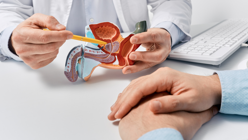
A raised prostate-specific antigen (PSA) level or an abnormal prostate MRI can understandably cause anxiety. If your urologist recommends a prostate biopsy, one of the first questions you may ask is, “What type of prostate biopsy should I have?”
Advances in medical imaging have changed the way prostate cancer is diagnosed. Today, many men undergo MRI-US fusion prostate biopsy, a modern technique that combines magnetic resonance imaging (MRI) with real-time ultrasound to improve the accuracy of prostate cancer detection.
Compared with the traditional random prostate biopsy, MRI-US fusion biopsy allows suspicious areas identified on MRI to be sampled more precisely. In addition, an increasing number of centres now perform prostate biopsies through the transperineal route, which offers a lower risk of infection than the traditional transrectal approach.
Understanding these advances can help you make a more informed decision if a prostate biopsy has been recommended.
Why Is a Prostate Biopsy Needed?
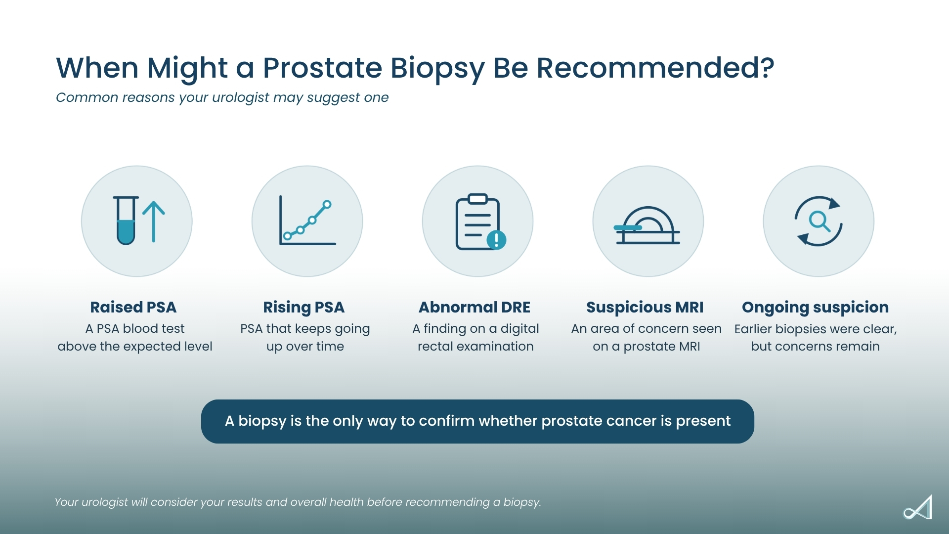
A prostate biopsy is the only way to confirm whether prostate cancer is present.
Your urologist may recommend a biopsy if you have:
- An elevated PSA blood test
- A rising PSA over time
- An abnormal digital rectal examination (DRE)
- A suspicious lesion seen on multiparametric MRI (mpMRI)
- Previous biopsies that were negative despite persistent suspicion of prostate cancer
The aim of a biopsy is not simply to detect cancer, but also to determine whether the cancer is clinically significant and requires treatment.
What Is a Standard Prostate Biopsy?
For many years, the standard method of diagnosing prostate cancer was the systematic or random template biopsy.
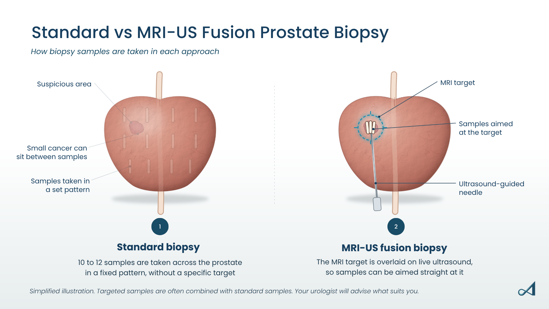
During this procedure, the urologist uses ultrasound guidance to take multiple biopsy samples from different regions of the prostate. Typically, 10 to 12 cores are obtained according to a standard template, regardless of whether a suspicious lesion is visible.
This approach increases the likelihood of finding cancer by sampling different parts of the prostate.
However, because the samples are taken systematically rather than directed at a specific abnormality, important cancers may occasionally be missed, while very small, low-risk cancers that may never cause problems can sometimes be detected.
What Is MRI-US Fusion Prostate Biopsy?
MRI-US fusion biopsy is an imaging-guided approach that combines multiparametric MRI (mpMRI) and real-time ultrasound.
Before the biopsy, the patient undergoes an MRI scan of the prostate. If the MRI identifies a suspicious area, the images are digitally fused with live ultrasound images during the biopsy procedure.
This allows the urologist to see the suspicious lesion in real time and guide the biopsy needle precisely into the target.
Rather than relying solely on random sampling, MRI-US fusion biopsy enables targeted sampling of the areas most likely to contain clinically significant prostate cancer.
Many centres also combine targeted biopsies with systematic template biopsies to support diagnostic accuracy.
MRI-US Fusion Biopsy vs Standard Random Biopsy
A potential benefit of MRI-US fusion biopsy is targeted sampling of suspicious areas.
Compared with standard random biopsy, MRI-targeted biopsy has been shown to:
- Improve detection of clinically significant prostate cancer
- Reduce the diagnosis of low-risk cancers that may not require treatment
- Target sampling of suspicious MRI lesions
- Support targeted sampling in men with previous negative biopsies
- Inform treatment planning
In other words, MRI-US fusion biopsy helps doctors identify clinically significant cancers while reducing unnecessary detection of indolent disease.
This is one approach to more personalised prostate cancer diagnosis.
How Does MRI-US Fusion Biopsy Compare in Accuracy?
Multiple international studies have demonstrated that MRI-targeted biopsy improves the detection of clinically significant prostate cancer compared with systematic biopsy alone.
For men with suspicious MRI findings, targeted biopsy is more likely to identify higher-grade cancers that require treatment.
However, MRI is not perfect.
Occasionally, clinically significant cancers may not be visible on MRI. For this reason, many urologists perform both targeted and systematic biopsies during the same procedure to support cancer detection.
Current international guidelines increasingly recommend pre-biopsy MRI before prostate biopsy whenever possible.
Transperineal vs Transrectal Prostate Biopsy: What’s the Difference?
Another important development in prostate biopsy is the route used to obtain the tissue samples.
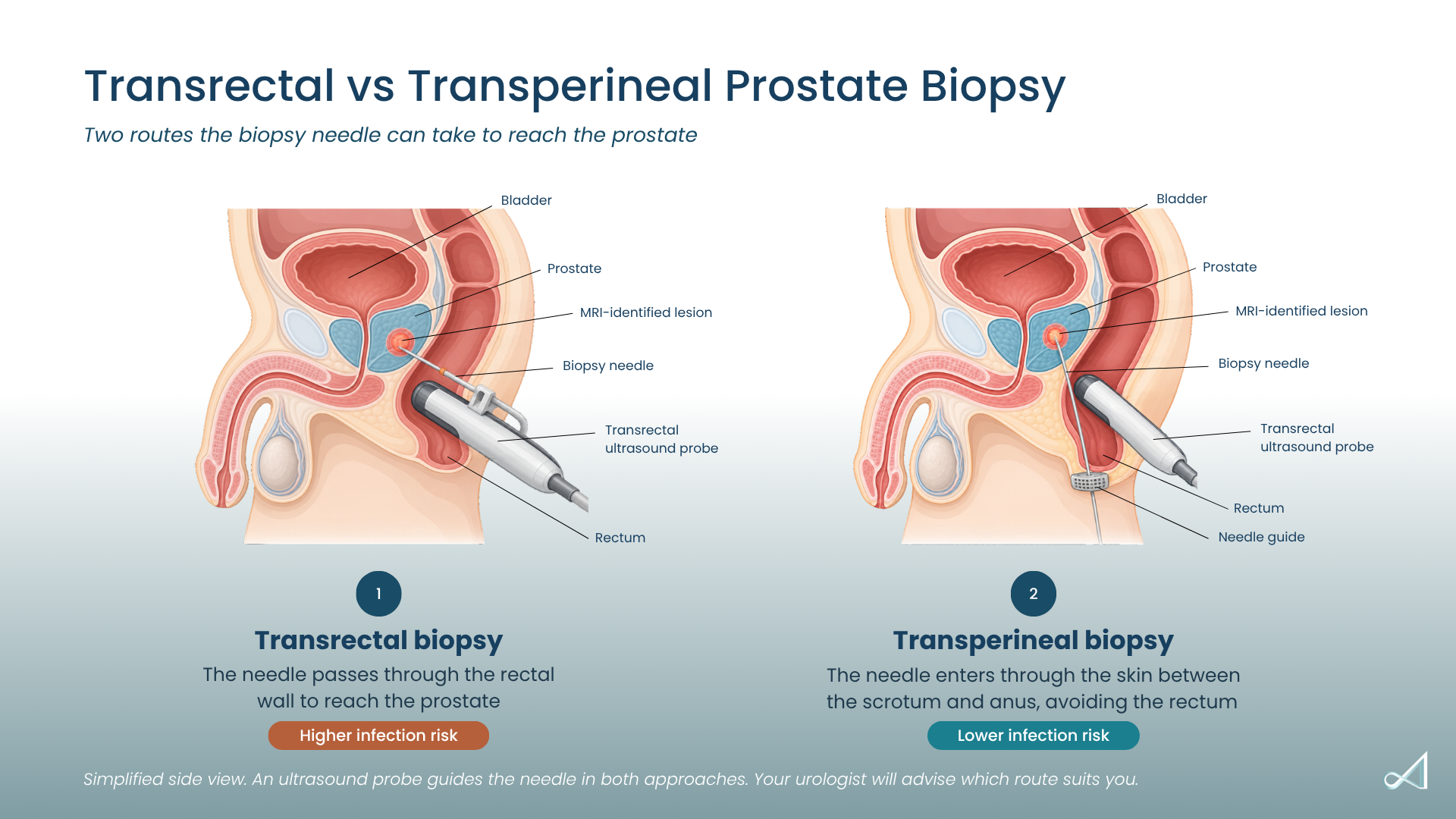
Traditionally, prostate biopsies were performed through the transrectal route, where the biopsy needle passes through the wall of the rectum into the prostate.
Today, some urologists have adopted the transperineal approach, where the biopsy needle passes through the skin between the scrotum and anus (the perineum).
Although both approaches can diagnose prostate cancer effectively, there are important differences.
Benefits of the Transperineal Prostate Biopsy
The transperineal approach has become increasingly popular because it is associated with a lower risk of infection.
Since the needle does not pass through the rectum, bacteria from the bowel are far less likely to enter the bloodstream or urinary tract.
Compared with transrectal biopsy, transperineal biopsy has several reported differences:
- Lower risk of serious infection and sepsis
- Reduced need for broad-spectrum antibiotics
- Access to the anterior regions of the prostate, where some cancers can be difficult to reach through the rectum
- Lower rates of antibiotic-resistant infections
- Sampling of the entire prostate gland
As antibiotic resistance continues to increase worldwide, reducing infection risk has become a major reason why many international guidelines now recommend the transperineal approach.
Are There Any Downsides to the Transperineal Approach?
Modern transperineal biopsy is generally well tolerated and is increasingly performed under local anaesthesia in outpatient settings.
Some patients may experience temporary:
- Blood in the urine
- Blood in the semen
- Mild bruising around the biopsy site
- Temporary discomfort when sitting
- Temporary difficulty passing urine
These side effects are usually mild and resolve within several days to weeks. Of note, these side effects are present in the transrectal approach too.
Serious complications are uncommon.
Is Transrectal Biopsy Still Used?
Yes.
Transrectal biopsy remains widely available and continues to diagnose prostate cancer effectively.
However, because of the higher risk of infection and increasing concerns regarding antibiotic-resistant bacteria, many urologists worldwide have increasingly adopted transperineal biopsy.
The choice often depends on the urologist’s expertise, equipment availability, and individual patient factors.
How Is the Prostate Biopsy Approach Chosen?
There is no single biopsy technique that is appropriate for every patient.
For many men, the combination of pre-biopsy MRI, MRI-US fusion targeted biopsy, and the transperineal approach is one diagnostic approach that may be considered.
This approach combines:
- Detection of clinically significant cancers
- Reduced detection of insignificant cancers
- MRI-guided targeting of suspicious areas
- Lower infection risk
- Sampling of different regions of the prostate
Your urologist will recommend the most appropriate biopsy technique based on your PSA level, MRI findings, prostate anatomy, previous biopsy history, and overall health.
The Bottom Line
The diagnosis of prostate cancer has changed over the past decade. MRI-US fusion prostate biopsy has improved the ability to detect clinically significant cancers by combining the detailed imaging of MRI with the precision of real-time ultrasound guidance. Compared with traditional random prostate biopsy, it allows suspicious lesions to be targeted more accurately while reducing unnecessary diagnosis of low-risk disease.
At the same time, the transperineal approach has been adopted in many centres because it offers a lower risk of infection than the traditional transrectal technique while providing access to all regions of the prostate.
If you have been advised to undergo a prostate biopsy, discussing whether MRI-US fusion biopsy and the transperineal approach are suitable for your situation can help you understand the potential diagnostic benefits and risks of each approach.
Frequently Asked Questions
How does MRI-US fusion biopsy compare with a standard prostate biopsy?
MRI-US fusion biopsy allows targeted sampling of suspicious areas identified on MRI, which may improve detection of clinically significant prostate cancer. Many urologists also perform systematic biopsies at the same time to maximise cancer detection.
How does infection risk compare between transperineal and transrectal biopsy?
Transperineal biopsy is associated with a lower risk of infection because the biopsy needle does not pass through the rectum. This can reduce the risk of serious infections such as sepsis.
Does MRI-US fusion biopsy replace the need for systematic biopsy?
Not always. While MRI-targeted biopsy improves the detection of significant cancers, many specialists perform both targeted and systematic biopsies during the same procedure to avoid missing cancers that are not visible on MRI.
Is a prostate biopsy painful?
Most prostate biopsies are performed under local anaesthesia or sedation. Patients may experience temporary discomfort, but the procedure is generally well tolerated and usually performed as a day surgery, and most men return home on the same day.
About Dr Terence Lim Sey Kiat
Dr Terence Lim (林诗杰医生) is a Senior Consultant Urologist (泌尿外科高级顾问) in Singapore sub-specialising in Uro-Oncology with a focus on prostate and kidney cancer surgeries. He is also the Medical Director at Assure Urology & Robotic Centre (安顺泌尿外科中心的医疗总监).
This article is for general education and does not replace a proper consultation. If you have concerns about your urological health, an assessment with a urologist is the best next step to understand your options.